Barcelona · International referrals
MD, PhD Cornea & Ocular Surface Keratoprosthesis Surgery

Restoring vision beyond transplantation.

Advanced keratoprosthesis and ocular surface reconstruction for patients with severe corneal blindness in whom conventional transplantation has a poor prognosis.
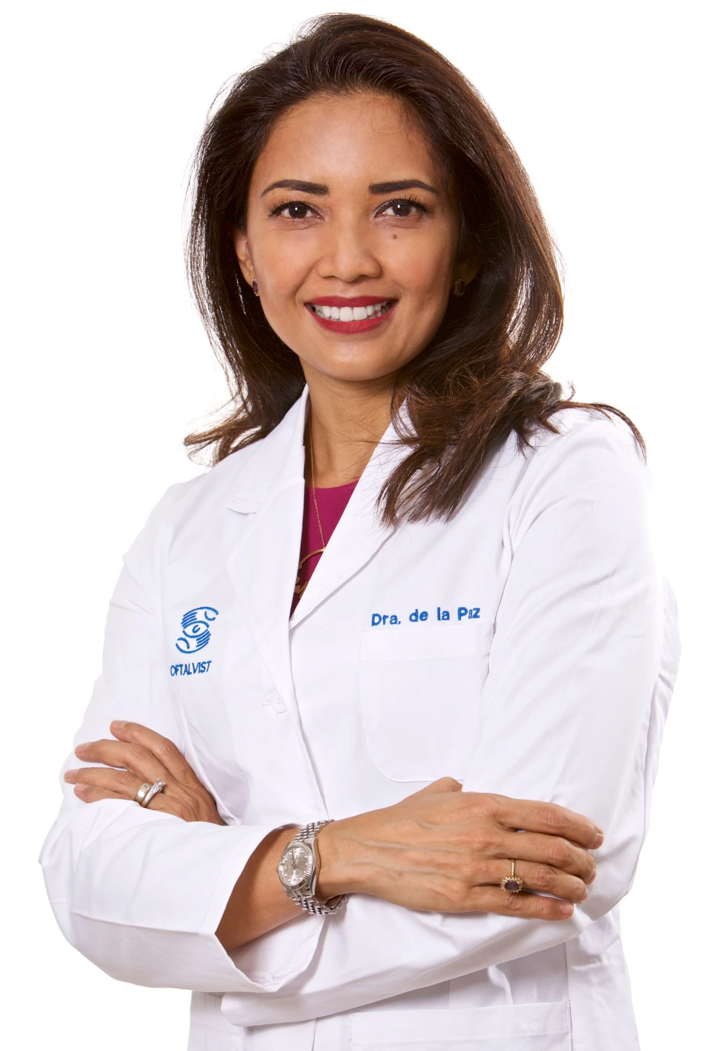
Dr. María Fideliz de la Paz
Boston KPro · OOKP · Tibial Bone KPro · Transmucosal Boston KPro
Boston KProArtificial cornea
OOKPOsteo-odonto
Tibial KProAutologous bone support
TransmucosalBuccal mucosa coverage
Advanced corneal reconstruction

When a donor cornea is no longer enough.

Repeated graft failure, severe chemical injury, autoimmune cicatrizing disease and extreme ocular-surface damage can make conventional keratoplasty unlikely to succeed.

The objective is not to apply the same prosthesis to every eye, but to select the reconstructive strategy that best matches the ocular surface, visual potential and long-term risks of each patient.

01

Boston Keratoprosthesis

Artificial corneal replacement for carefully selected eyes with a poor prognosis for conventional grafting and an ocular surface able to support a Boston KPro strategy.

02

Osteo-Odonto-Keratoprosthesis

A highly specialized staged procedure using autologous tooth and alveolar bone as biological support for the optical cylinder in severe end-stage ocular surface disease.

03

Tibial Bone Keratoprosthesis

A biological-support alternative using autologous tibial bone in complex cases where an osteo-dental lamina cannot be used or is not the preferred option.

04

Boston KPro + Buccal Mucosa

Combined reconstructive approaches integrating Boston keratoprosthesis with autologous oral mucosa for eyes with severe ocular surface compromise.

Could keratoprosthesis be an option? Request an initial evaluation in Barcelona or an online consultation before travelling.
Evaluate my case online Book in Barcelona
Technique focus

Boston Keratoprosthesis Type I

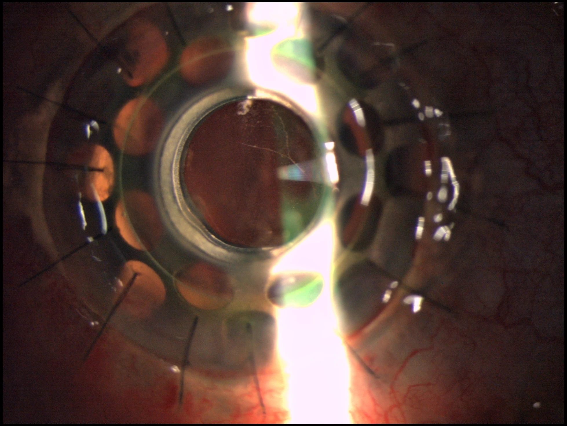
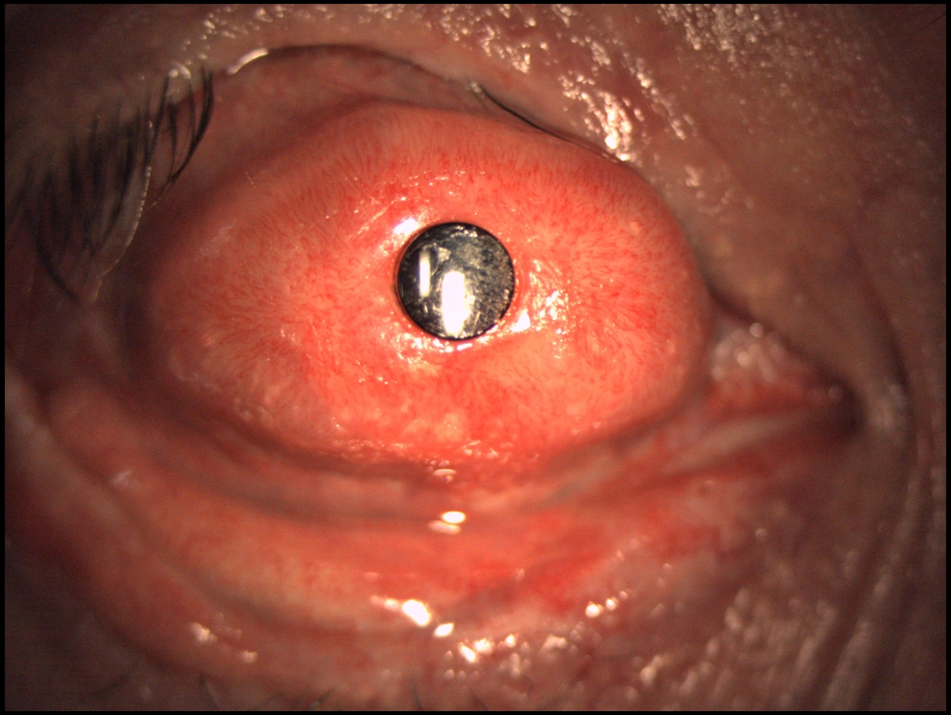
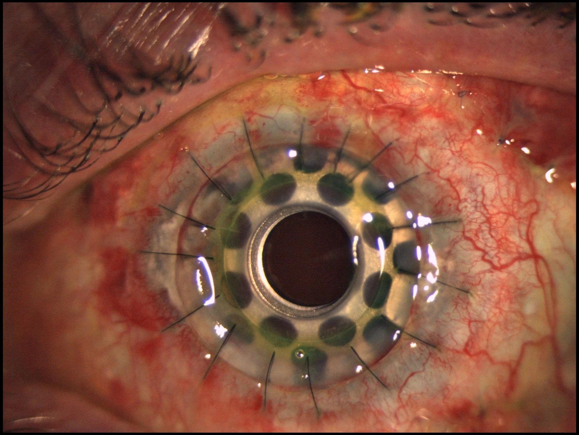
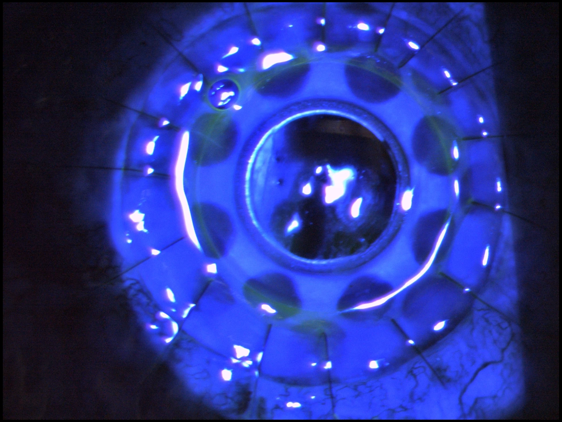
The Boston Type I Keratoprosthesis is one of the most established artificial cornea systems for eyes in which conventional corneal transplantation has a high risk of failure but the ocular surface remains capable of supporting a Type I approach.

The aim of this section is to show the device in a clinical, real-world manner: not as an abstract diagram, but as a surgical solution seen at the slit lamp in actual postoperative eyes.
CE marked for the European market Boston Keratoprosthesis Type I received European Conformity (CE) marking in 2014, confirming conformity with the applicable European requirements at the time of certification. Regulatory information · NICE ↗
Indications
High-risk corneal blindnessRepeated graft failure, selected chemical injuries and ocular surface conditions where a donor cornea has a low chance of long-term clarity.
Strategy
Not every severe eye needs OOKPWhen the surface is suitable, Boston KPro may provide a less complex reconstructive pathway than osteo-odonto or tibial bone techniques.
Follow-up
Long-term surveillance is essentialSurface stability, epithelial status, glaucoma and posterior segment monitoring remain critical after surgery.
Have you been told that another corneal graft is unlikely to succeed? Send your reports, corneal photographs and previous surgical history for a preliminary online review.
Request online case review
Technique focus

Osteo-Odonto-Keratoprosthesis (OOKP)

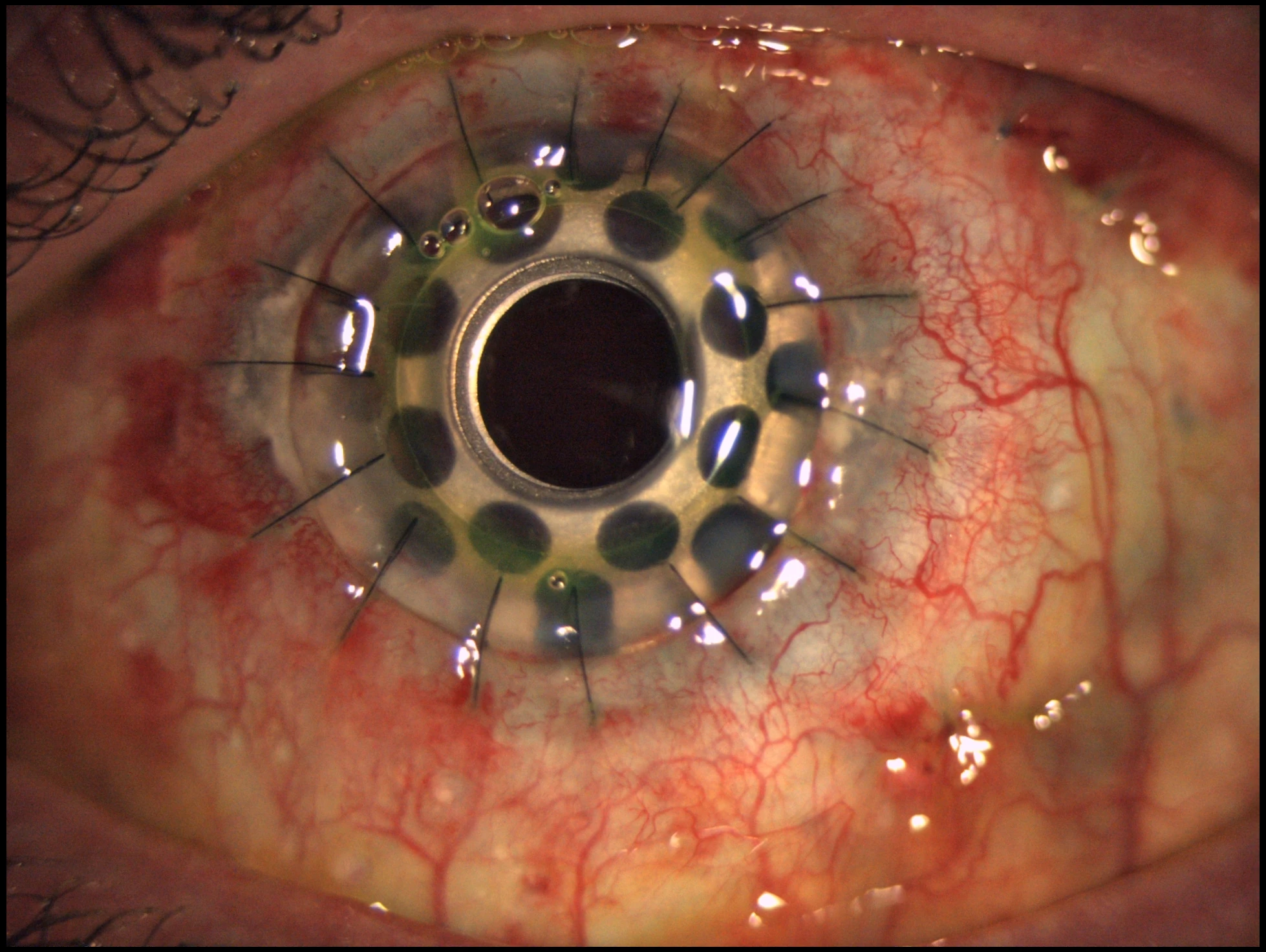
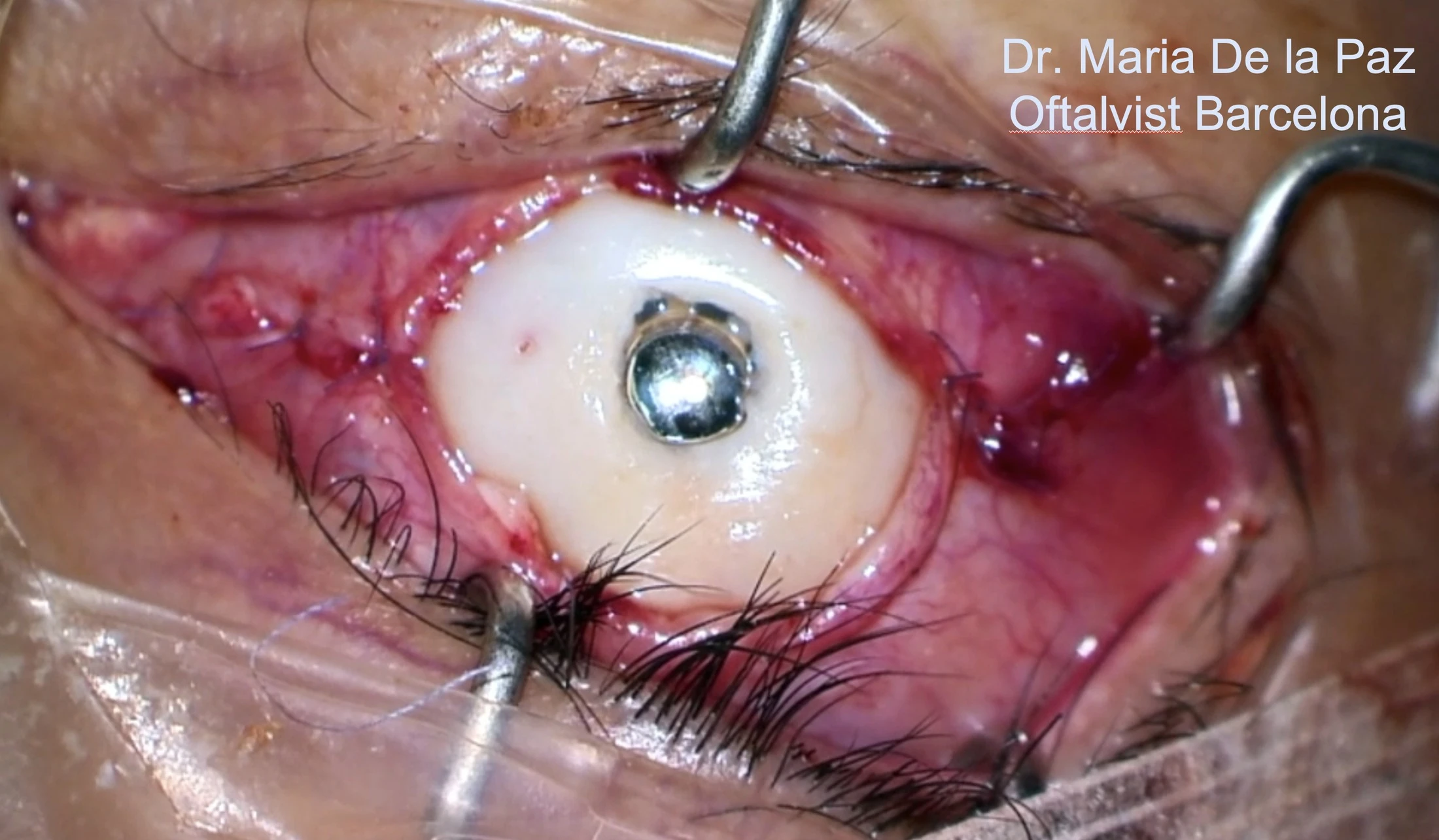
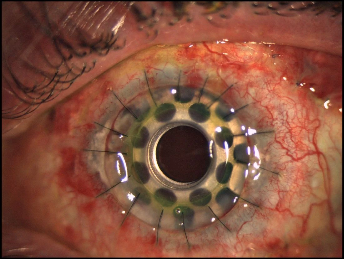
OOKP is one of the most complex and remarkable reconstructive procedures in ophthalmology. It is designed for end-stage ocular surface disease, especially in severely dry, keratinized or cicatrizing eyes in which a conventional corneal graft or even a standard Type I keratoprosthesis is unlikely to survive.

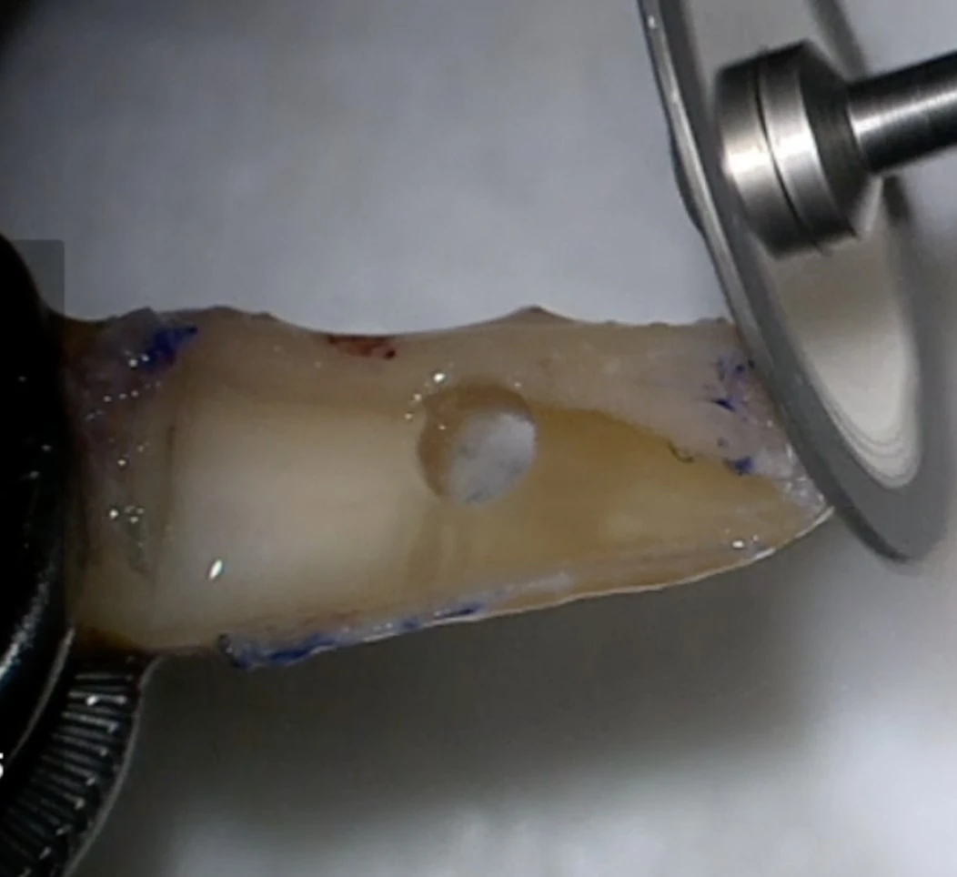
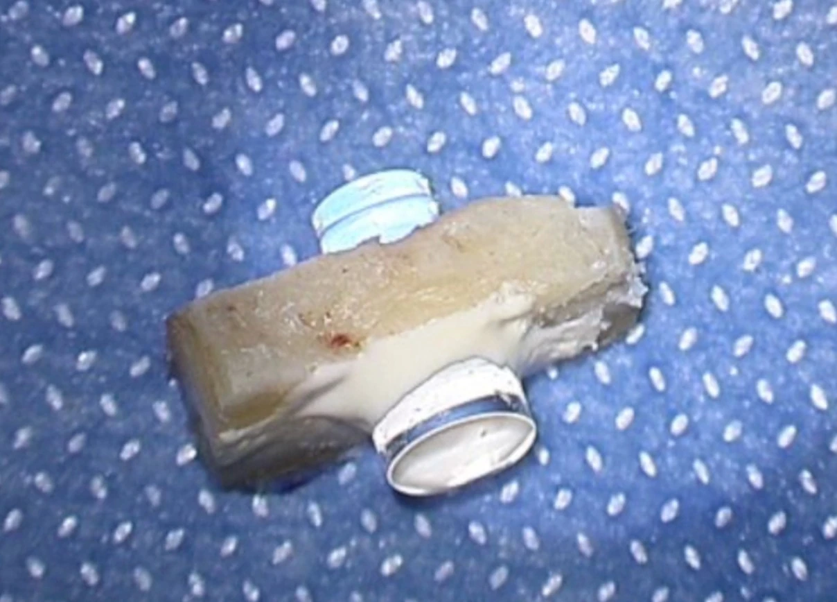
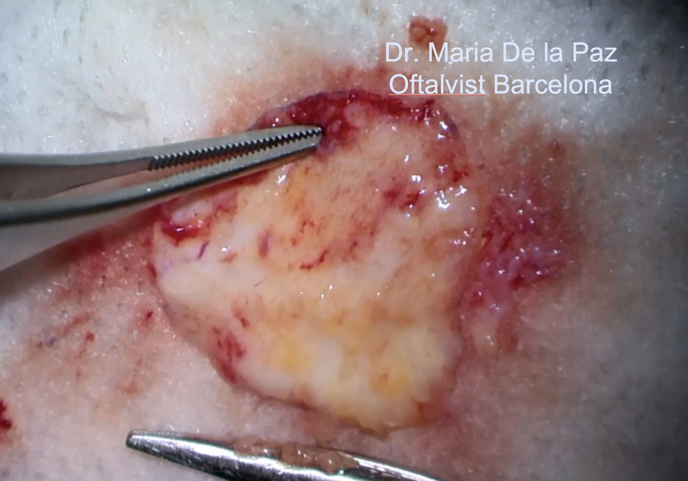
The technique uses an autologous tooth-bone lamina to support the optical cylinder and is usually combined with mucosal surface reconstruction, creating a biological environment capable of protecting the device in the most hostile ocular surfaces.

Indications
End-stage dry ocular surface diseaseSevere chemical burns, cicatrizing disease, keratinization and advanced surface failure where more conventional options are not appropriate.
Concept
Biological support for an optical cylinderThe osteo-dental lamina acts as a living autologous support, making OOKP fundamentally different from Boston Type I keratoprosthesis.
Selection
A surgery for very specific eyesPatient selection, systemic history and visual potential assessment are essential before indicating OOKP.
Current mockup update: this section now uses your real OOKP photographs, which immediately gives the website more authority and makes the distinction between Boston KPro and OOKP much clearer for both patients and referring ophthalmologists.
Severe dry, keratinized or cicatrizing ocular surface? OOKP candidates require detailed selection. An online consultation can determine whether a full in-person assessment is appropriate.
Request OOKP evaluation
Technique focus

Tibial Bone Keratoprosthesis

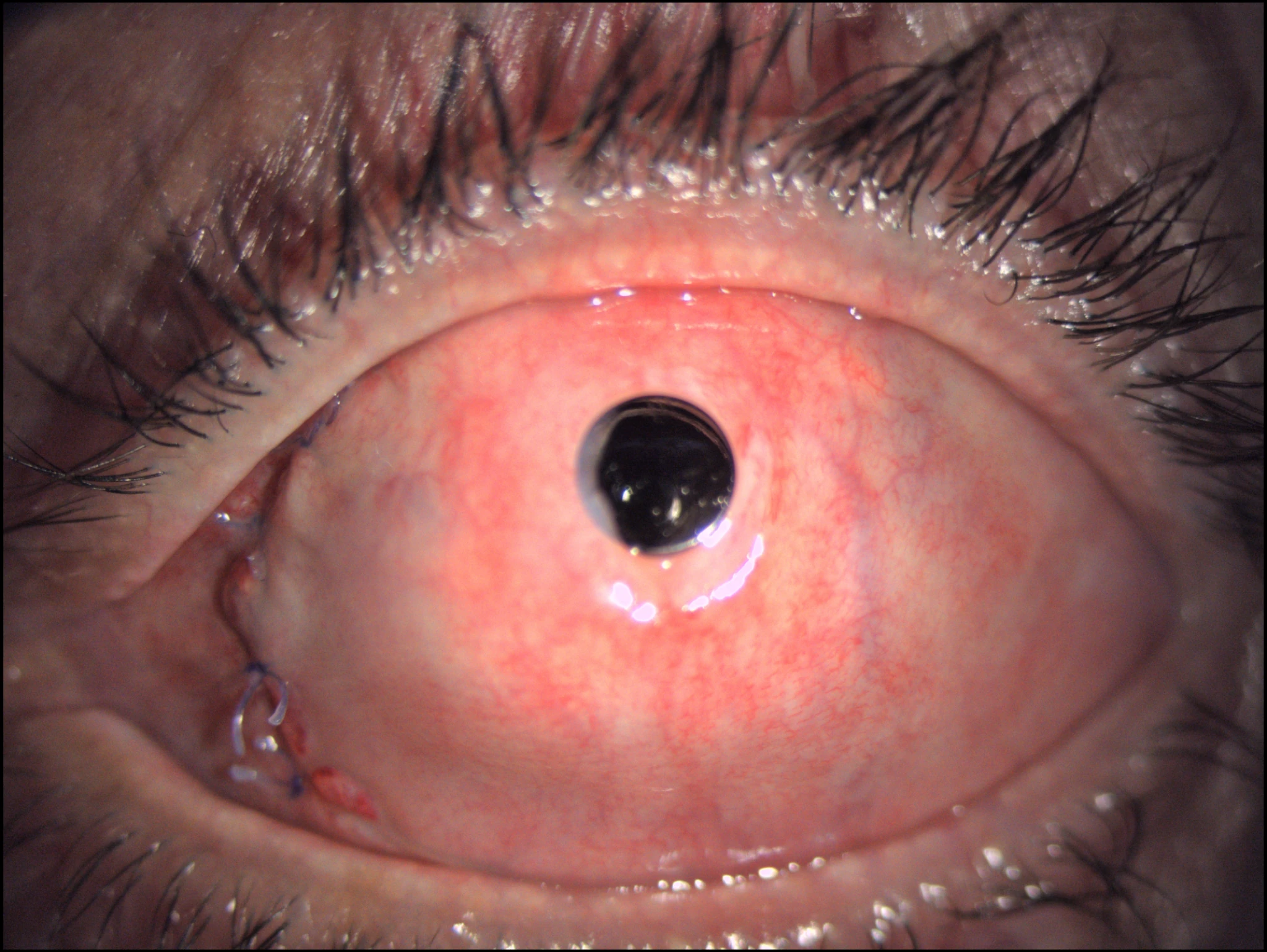
Tibial Bone Keratoprosthesis uses autologous tibial bone as the biological support for the optical cylinder. It is an alternative to osteo-odonto-keratoprosthesis in highly complex ocular-surface disease, especially when a tooth-bone lamina is not available or not ideal.

This technique belongs to the group of autologous-support keratoprostheses and is intended for very specific end-stage eyes. Its value lies in expanding the reconstructive options available when more conventional corneal procedures are no longer appropriate.
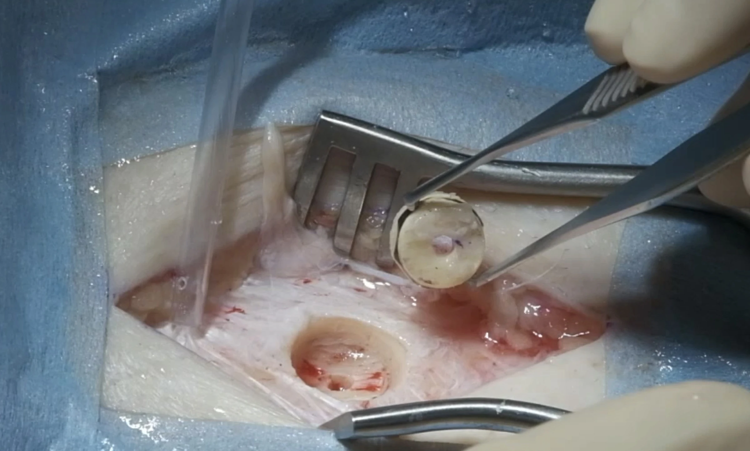
Role
Alternative biological supportTibial bone provides a structural autologous support when an osteo-dental lamina cannot be used.
Context
Part of the advanced KPro spectrumIt should be understood alongside Boston KPro and OOKP, not as a replacement for all cases but as a highly selective reconstructive option.
Selection
Reserved for complex indicationsAppropriate evaluation of ocular surface status, visual potential and the feasibility of other KPro strategies is essential.
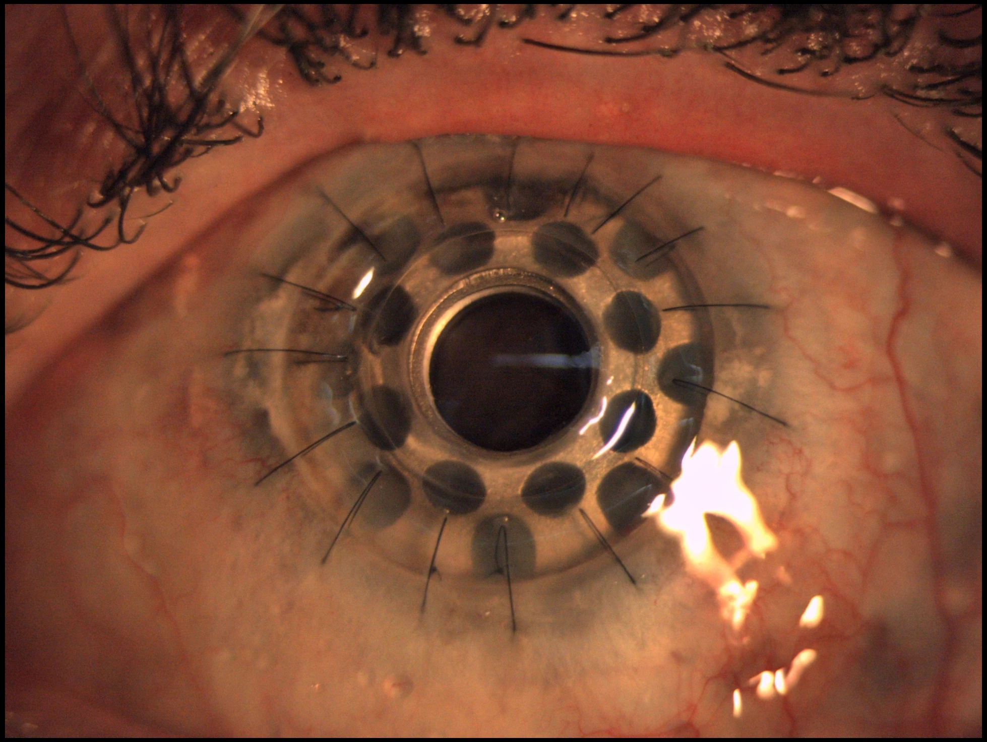
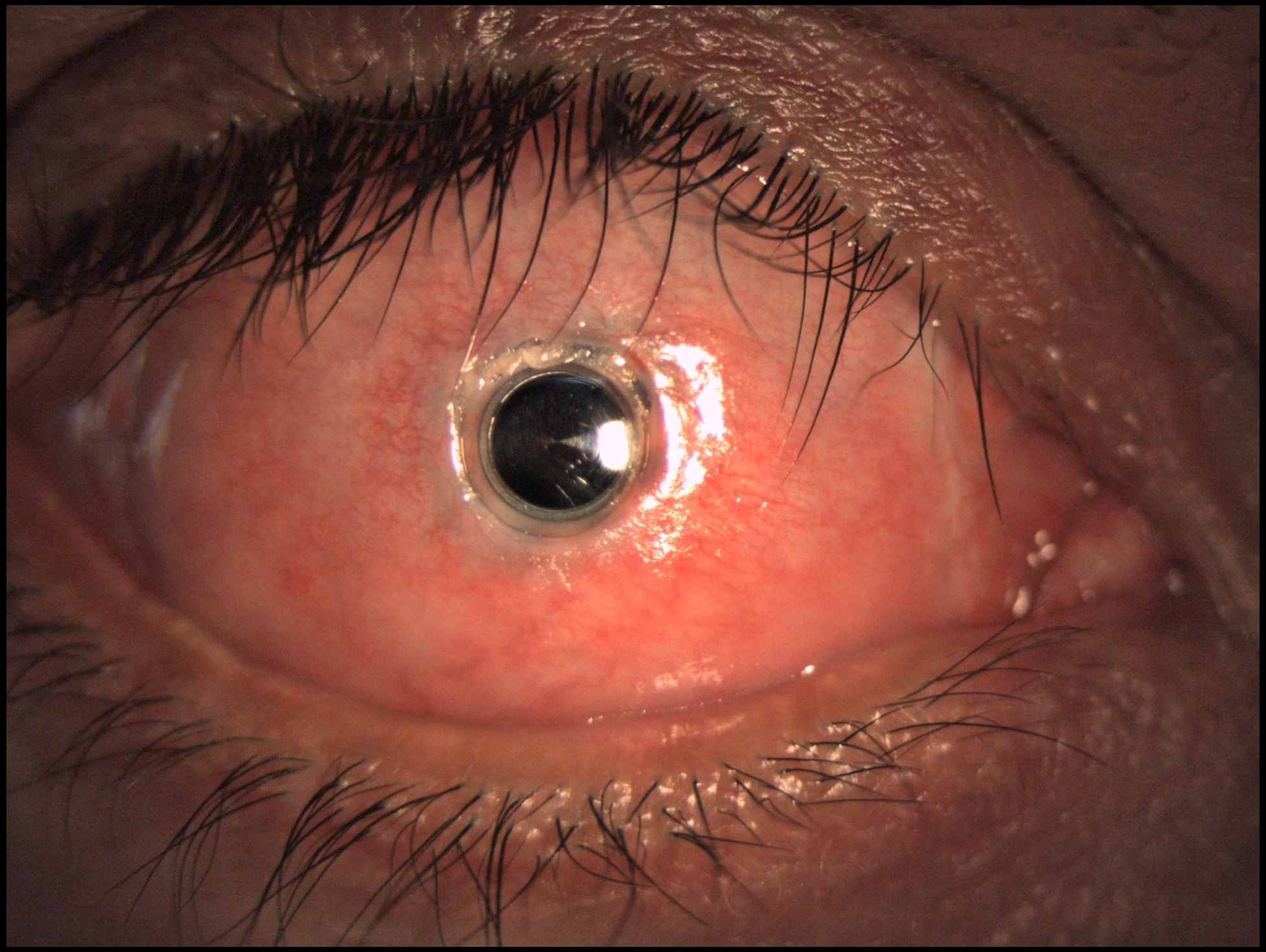
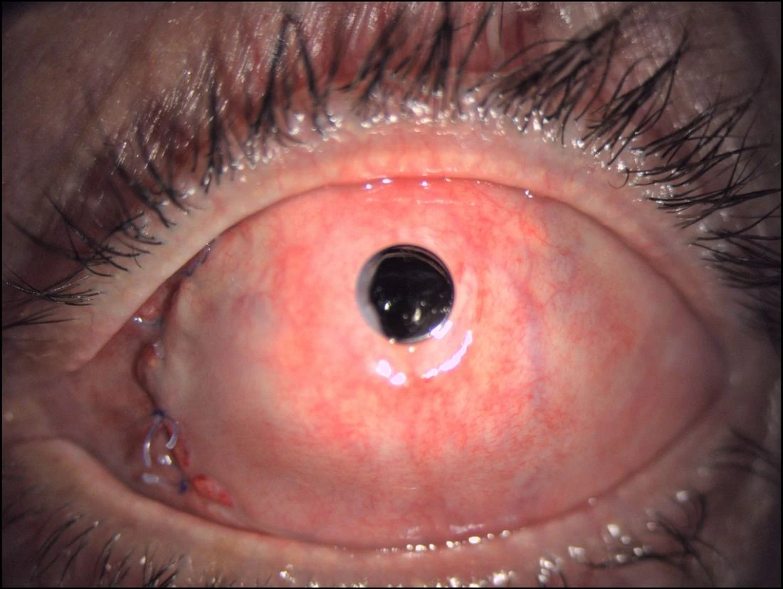
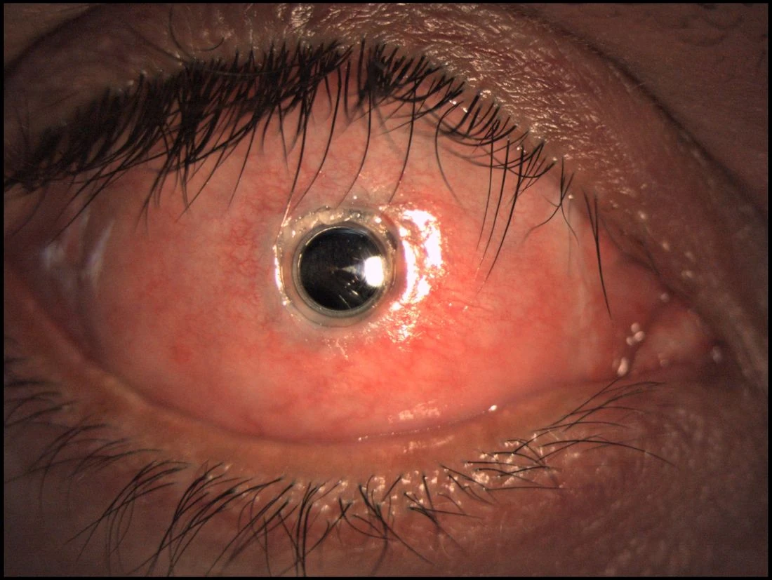
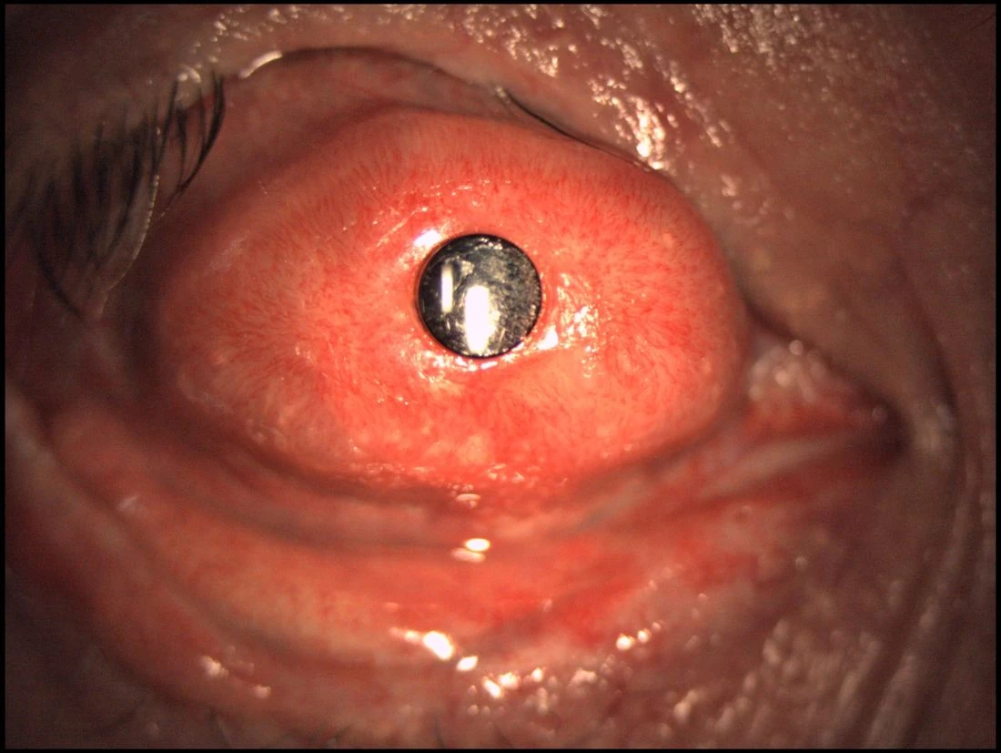
Current mockup update: this section now includes your real Tibial KPro clinical images, so the website already presents the three major categories of keratoprosthesis with authentic visual material.
Tibial Bone Keratoprosthesis frontal view
Tibial Bone KPro · Frontal clinical view
Tibial Bone Keratoprosthesis clinical photograph
Tibial Bone KPro · Clinical detail
Need a second opinion on OOKP versus Tibial KPro? The case can be reviewed remotely before arranging a trip to Barcelona.
Request a second opinion
Technique focus

Boston Keratoprosthesis with Buccal Mucosa

Transmucosal Boston keratoprosthesis combines the optical concept of Boston KPro with the protective and reconstructive role of autologous oral mucosa. This strategy is particularly relevant in eyes with advanced ocular-surface alteration where a standard Type I environment is not sufficient.

It represents an intermediate and highly specialized reconstructive pathway between conventional Boston KPro and the more complex autologous-support procedures such as OOKP or tibial bone keratoprosthesis.

Indications
Severe ocular-surface compromiseUseful in selected eyes with significant surface alteration in which mucosal coverage can improve long-term protection and integration of the device.
Concept
Boston optics + mucosal reconstructionThe procedure merges prosthetic corneal rehabilitation with surface reconstruction using autologous buccal mucosa.
Value
Expands the range of Boston-based solutionsIt widens the therapeutic spectrum for eyes that fall between standard Boston KPro and full autologous-support keratoprosthesis strategies.
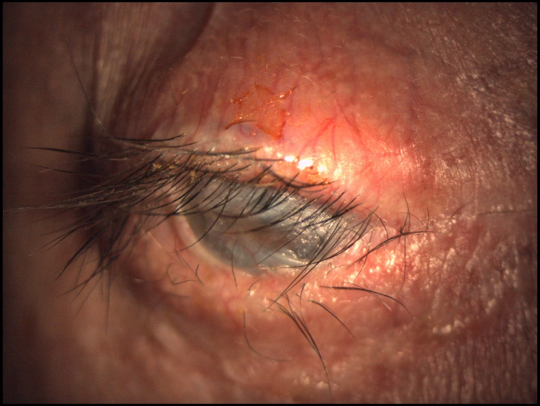
Current mockup update: the website now includes real clinical images of Boston KPro with buccal mucosa, completing the four principal keratoprosthesis categories presented by Dr. de la Paz.
Complex ocular-surface case? Request a specialist assessment to determine whether Boston Type I, OOKP, Tibial KPro or a transmucosal Boston strategy may be considered.
Send case for evaluation
Clinical decision framework

Which Keratoprosthesis?

The first decision is not “which device?”, but whether keratoprosthesis is appropriate at all. In Dr. de la Paz's clinical framework, the eye must represent end-stage ocular-surface disease with useful retinal and optic-nerve potential. The ocular surface phenotype then becomes the key determinant of the prosthetic strategy.

Gate 1 · Is there realistic visual potential?

Functional retina and optic nerve are prerequisites before undertaking end-stage ocular-surface reconstruction.

Patient selection precedes device selection.
Wet blinking eye

Preserved surface environment

  • Complete blink
  • No significant keratinization
  • Complete lid closure
  • Inflammation under control
Primary KPro pathway → Boston KPro Type I
Dry non-blinking eye

Severely cicatrized surface

  • Severe ocular-surface fibrosis
  • Symblepharon / ankyloblepharon
  • Keratinization
  • Severe dry eye or poor lid closure
Advanced reconstructive pathway → OOKP · Tibial KPro · Boston Type II / transmucosal strategy
High riskAutoimmune disease: SJS, ocular cicatricial pemphigoid, Mooren ulcer, Sjögren syndrome.
Intermediate riskSevere chemical injury.
Lower-risk KPro groupRepeated graft failure, aniridia, silicone-oil keratopathy, LSCD, herpetic opacity and other non-autoimmune causes.
Side-by-side

Four reconstructive strategies

These categories are not interchangeable. Surface wetness, blinking and keratinization, together with the underlying disease and visual potential, determine the direction of treatment.
Boston KPro Type I
Wet blinking eye

Boston KPro Type I

Best surface profileComplete blink, no keratin, complete lid closure, controlled inflammation.
Main roleFirst prosthetic category considered when a wet ocular surface can support Type I KPro.
Relative burdenLess reconstructively complex than autologous-support KPro, but lifelong surveillance remains essential.
Key surveillanceGlaucoma, infection, posterior segment and device/surface complications.
Osteo-Odonto-Keratoprosthesis
Dry non-blinking eye

OOKP

SupportAutologous tooth-bone lamina supporting an acrylic optic.
Main strengthTrue biological integration with minimal intraocular foreign material; systemic immunosuppression is not mandatory.
Trade-offsVery steep learning curve, smaller visual field, cosmetic limitations, difficult reversibility and difficult repetition.
Key surveillanceMucosal problems, extrusion, bone resorption, infection, glaucoma and posterior-segment complications.
Tibial Bone Keratoprosthesis
Dry non-blinking eye

Tibial Bone KPro

SupportAutologous tibial bone supporting the optical cylinder.
Main roleBiological-support KPro in the same severe dry-eye spectrum as OOKP.
Clinical contextUseful within the autologous-support pathway when the reconstructive plan favors tibial bone rather than osteo-dental support.
Key surveillanceMucosal complications, extrusion, bone resorption, infection, glaucoma, RD/CME and vitritis.
Boston KPro with buccal mucosa
Severely altered surface

Boston KPro + Buccal Mucosa

ConceptBoston optics combined with mucosal surface reconstruction.
Clinical nicheSelected severely altered ocular surfaces requiring mucosal protection rather than a standard Type I environment.
Presentation contextDr. de la Paz places Boston Type II among options for dry non-blinking eyes; her published work also includes transmucosal Boston approaches.
Key pointA hybrid reconstructive option rather than a simple extension of standard Boston Type I.
Simplified referral algorithm

From ocular surface to KPro family

01 End-stage ocular surface disease? High graft-rejection risk, repeated graft failure, LSCD, severe burns, cicatrizing disease or failure of prior surface-reconstruction procedures.
02 Retina and optic nerve functional? If visual potential is inadequate, the rationale for a high-risk KPro procedure changes fundamentally.
03 Wet blinking or dry non-blinking? Wet → Boston Type I pathway. Dry/cicatrized → OOKP, Tibial KPro or selected transmucosal / Type II Boston strategy.

This is an educational referral framework, not an automatic surgical indication. Final choice requires a complete ocular-surface, glaucoma, retinal and systemic assessment.

International consultation

Not sure which keratoprosthesis fits the case?

A preliminary medical evaluation can be performed online. If surgery appears potentially appropriate, a complete in-person assessment can then be arranged in Barcelona.
Request online consultation
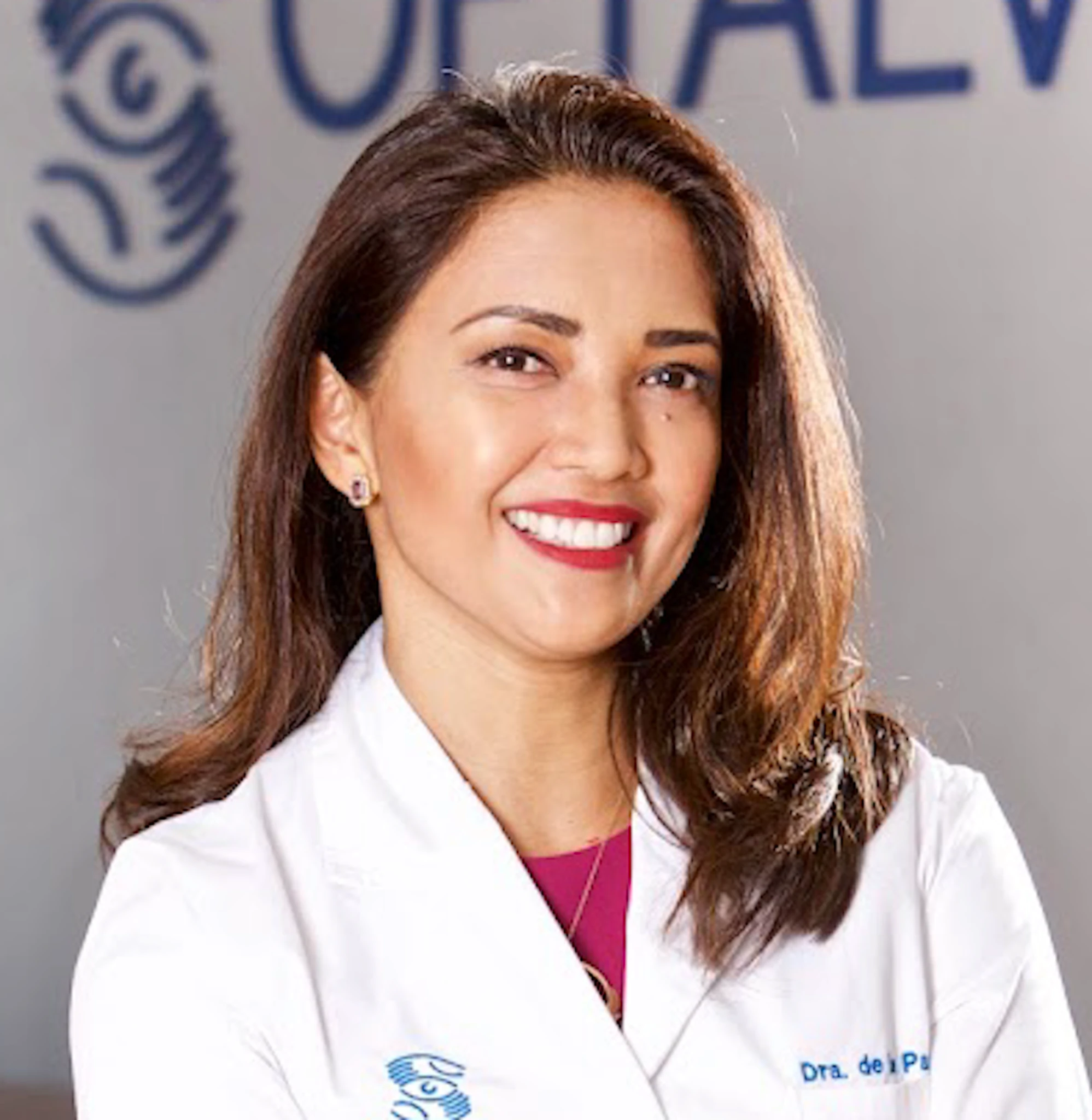
Dr. María Fideliz de la Paz
The surgeon

Dr. María Fideliz de la Paz

Advanced corneal blindness requires a strategy beyond conventional transplantation.

Dr. de la Paz is an anterior segment, cornea and ocular surface surgeon based in Barcelona. Her clinical work includes limbal transplantation, ocular surface reconstruction, penetrating keratoplasty, Boston keratoprosthesis, osteo-odonto-keratoprosthesis and tibial bone keratoprosthesis.

Doctoral DegreeUniversitat Autònoma de Barcelona · 2016
Distinguished KPro SurgeonMassachusetts Eye and Ear / Harvard · 2019
Oftalvist BarcelonaCornea, ocular surface & anterior segment
International PracticeThe View Hospital · Doha, QatarBurjeel Medical Center · Abu Dhabi, UAE
2001–03Master in Anterior Segment, Institut Universitari Barraquer, Barcelona
2004–07Fellowship in Anterior Segment, Centro de Oftalmología Barraquer
2008–22Consultant Specialist in Cornea, External Eye Disease, Ocular Surface and Eye Banking, Barraquer Eye Center
2022–Cornea, ocular surface and anterior segment practice at Oftalvist Barcelona
Patient pathway

Which keratoprosthesis?

01
Previous corneal historyFailed grafts, chemical burns, autoimmune disease, severe vascularization and previous ocular surgery.
02
Ocular surface assessmentTear film, dryness, fornices, conjunctiva, limbal status, keratinization and degree of cicatrization.
03
Visual potentialRetina, optic nerve, glaucoma and electrophysiological assessment when required.
04
Individualized techniqueBoston KPro, OOKP, tibial bone support or a transmucosal Boston KPro approach.
International profile

Teaching, collaboration and KPro surgery worldwide.

International surgical activity and invited lectures reinforce the site as a professional referral platform rather than a conventional clinic website.

2017–18
Invited surgeon, Philippine Cornea SocietyParticipation in the first osteo-odonto-keratoprosthesis performed at St. Luke's Medical Center.
2019
Distinguished Keratoprosthesis SurgeonMassachusetts Eye and Ear Infirmary / Harvard University.
2024
KPro Summit · Lisbon33rd Biennial Cornea Conference.
2025
Advanced Keratoprosthesis Symposium · BeijingInternational keratoprosthesis and keratoplasty meetings.
Appointments · Barcelona & Online

Request an evaluation.

Patients and referring ophthalmologists can request an in-person consultation in Barcelona or an online preliminary evaluation of the case. Clinical reports, slit-lamp photographs, imaging and previous surgical history can be reviewed before travel.

International Department
[email protected]
Oftalvist Barcelona
C/ Anglí, 40
08017 Barcelona · Spain
HLA Internacional Barcelona · 4th floor
Please indicate whether you are requesting an in-person appointment or an online case evaluation.
Online evaluation In-person visit